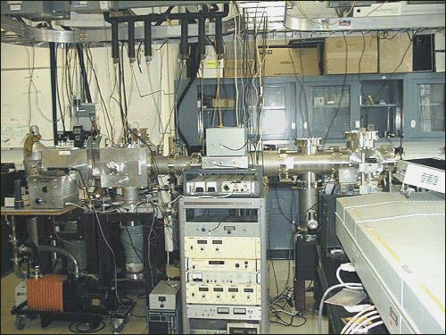
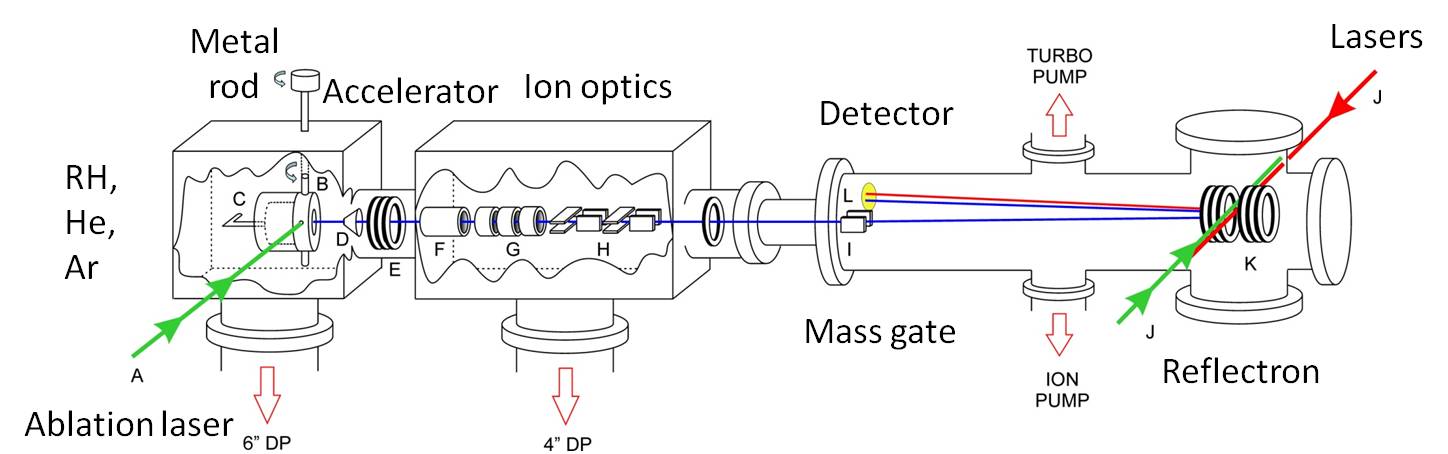
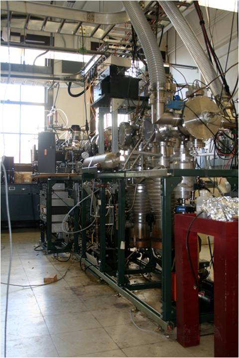
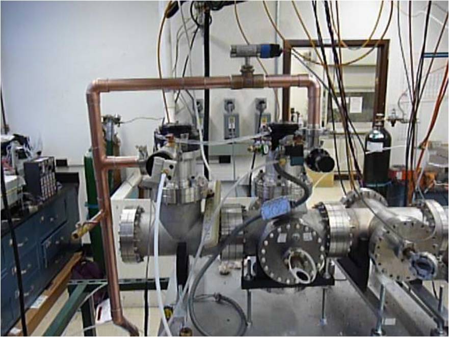
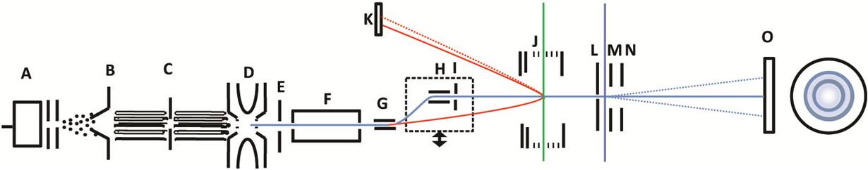
- Ions are formed by laser ablation, electrospray, or electric discharge (shown)
In the electric discharge source, a gas is introduced through a pulsed valve (A) and travels through charged plates.
The resulting electric discharge produces ions. The ions expand into vacuum and cool.
A skimmer (B) selects the central portion of the ion beam.
- Ions are collected and thermalized
The ions travel through octopole ion guides (C) and into a radio-frequency ion trap (D), where they are thermalized by
collisions with helium gas.
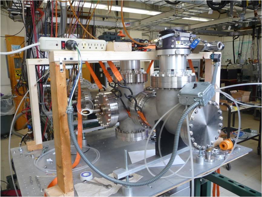
- Ions are extracted and mass selected
Ions are extracted and accelerated in a collinear Wiley-McLaren configuration (E) and
re-referenced to ground potential (F).
- Electronic Spectroscopy
A mass gate (G) deflects selected ions (shown in red) into the reflectron (J).At the turning point of the reflectron,
a tunable laser, in the visible, or UV dissociates the ions.
The masses of resulting ions are determined by their flight times to a microchannel plate detector (K).
- Photofragment Imaging Studies
A mass gate (G,H) deflects selected ions (shown in blue) through the reflectron (J) which is now grounded.
Irises (I,L) collimate the ion beam. A tunable laser in the visible or UV dissociates the ions.
Fragment ions are accelerated in a velocity map imaging configuration (L,M,N) and are collected on a gated imaging detector (O).
The resulting image (far right) is collected by a CCD camera.
|