Complementing the existing structural biology facilities, the newly opened X-ray diffraction facility allows researchers to solve macromolecular structures by X-ray crystallography. The facility consists of an RigakuMSC RU-H3R X-ray generator and RAXIS 4++ X-ray detector equipped with Osmic Confocal Max-Flux optics, an inverse phi goniostat and an Oxford cryojet. With the support of two crystallographic laboratories in the biochemistry department (Dr. Theis, Dr. Garman) and one in the chemistry department (Dr. Hardy), the facility is able to provide training, technical expertise, instrumentation and computational infrastructure for obtaining well-diffracting crystals, mounting crystals, measuring high-quality diffraction data, processing data, data backup, solving the structures by molecular replacement or MIR/MAD techniques. For example, users have access to a dynamic light scattering instrument housed in Dr. Theis’s laboratory for screening samples for crystallizability.
Contact: Karsten Theis, theis@biochem.umass.edu, 7-2890
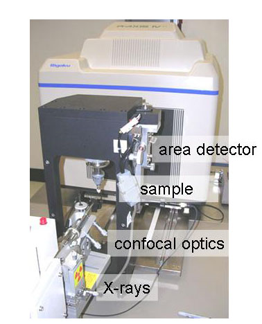
View of the optical bench of the X-ray diffraction facility. X-rays are produced by a rotating anode in the enclosure seen front left, collimated and focused by confocal optics to produce a bright X-ray beam hitting the sample (center). Diffracted X-rays are measured by the area detector on the sled in the background.
There is an experimental and a computational aspect to solving the structures of biomolecules:
Experiment: Measuring diffraction data from a single crystal
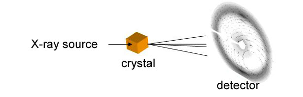
Computation: Calculating electron density from phases and measured intensities

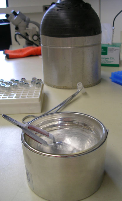
The facility provides equipment to freeze crystals in liquid nitrogen, to store them, measure them and ship them in the cryocooled state
 ........
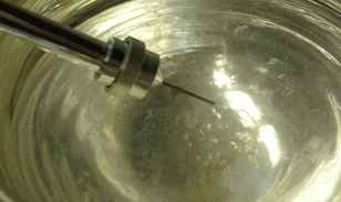
Left: Tools for mounting crystals on loops and freezing them. Right: A cryoloop attached to a magnetic wand is submerged in liquid nitrogen,
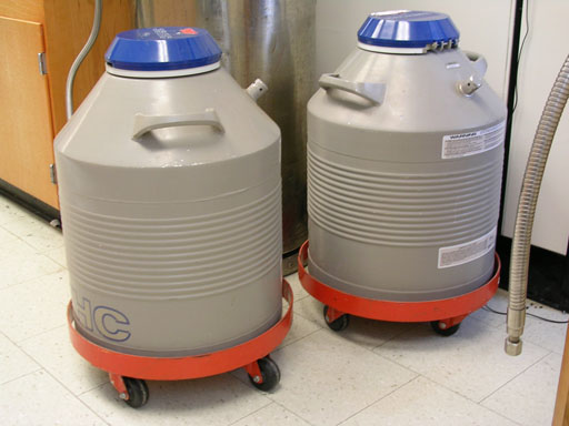
Dewars with a capacity to store up to 1200 crystals
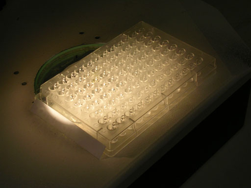
Crystal setups are stored and monitored in a dedicated crystallization room. Experiments may be performed at room temperature or 4, 15, or 37 degrees Celsius. Three stereomicroscopes are available for viewing and mounting crystals. Crystallization supplies and reagents are available at cost.
 ....................
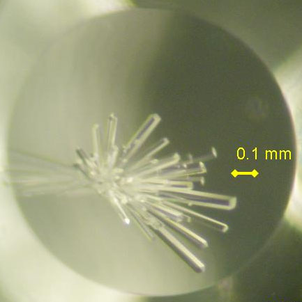
Crystallization setups viewed under the microscope